
Adelaide's fundamental pelvic floor imaging service
Our key sonographer introduced pelvic floor ultrasound services to Adelaide over 12 years ago and brings a wealth of unique knowledge to the clinic. We work closely with pelvic floor physiotherapists, gynaecologists and urogynecologists to support accurate diagnosis and targeted treatment.
Supporting Your Pelvic Health with Advanced Ultrasound Services
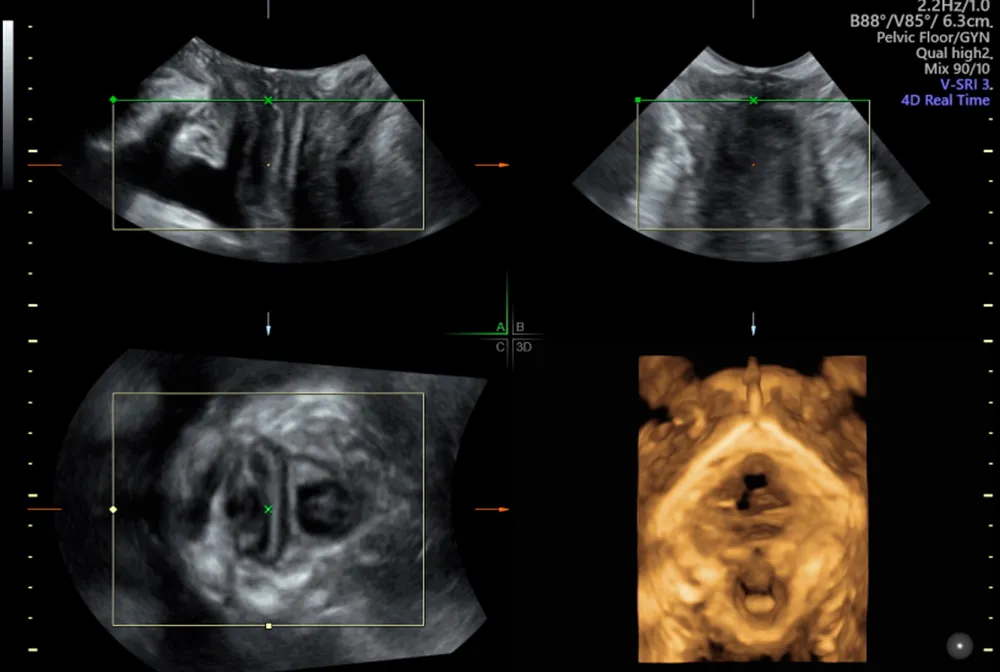
3D/4D Pelvic floor ultrasound is a study that can obtain images comparable to an MRI, with the advantage of watching the way the pelvic floor muscles are moving in real time. The data obtained can then be manipulated to view the pelvic floor muscles and organs in three planes and in multiple slices.
- Symptoms of voiding dysfunction
- Faecal incontinence
- Anal sphincter trauma
- Urinary stress and urge incontinence
- Pelvic muscle trauma post-delivery
- Post-surgical complications or recurrence associated with prolapse
- Assessment of mesh repair and TVT slings
- Symptoms of pelvic organ prolapse (sensation of lump or dragging)
- Symptoms of posterior compartment prolapse (straining at stool, chronic constipation, feeling of incomplete bowel emptying)
How Can an Ultrasound Help?

Can predict your risk of prolapse in the future
Measuring the pelvic floor outlet (levator hiatus) for any signs of ballooning can help predict post surgical prolapse recurrence. In addition, puborectalis avulsion can also impact prolapse recurrence. Both of these measures can assist your surgeon with the most appropriate surgical technique for your repair.
Helps with symptoms of urinary incontinence
With ultrasound we can look at bladder neck mobility. Over-rotation of the urethra often indicates stress incontinence. We can also look for funnelling of the bladder neck which is often associated with leakage or urge incontinence.
Determines pelvic floor muscle integrity
Determines pelvic floor muscle integrity post vaginal delivery for an informed management plan with your physiotherapist and obstetrician if planning further pregnancy.
Assists in diagnosing prolapse
A pelvic floor ultrasound can help quantify the degree of prolapse of either the bladder, uterus or rectum. It is especially useful in differentiating between different forms of posterior compartment problems.


Schedule Your Pelvic Floor Ultrasound Today
We are here to provide clarity, confidence, and exceptional care.
Your Trusted Partner in Advanced Women's Imaging
Our team of imaging specialists brings together years of experience and specialised training to provide you and your healthcare provider with clarity and exceptional detail.
We are committed to providing you with a supportive and understanding environment. Our team uses the latest ultrasound technology and advanced techniques to deliver detailed images, ensuring a thorough evaluation of your gynaecological health.

Why Patients and Providers
Choose Us for Pelvic Floor Ultrasound Services
Detailed Reports and Concise Results
We provide referrers with comprehensive reports that deliver clear, clinically meaningful results to support decision-making.
Compassionate, Patient-centred Care
Receive a personalised service from a small team of experienced professionals, ensuring attentive care for every patient.
Trained team
Rely on a team recognised for its extensive experience and significant contributions to medical research and education.
Advanced Diagnostics for Complex Cases
We provide detailed ultrasounds and clarification through second opinions for precise and trustworthy diagnostics.
Specialised Women’s AND Children’s Health Care
Benefit from pioneering expertise in endometriosis, paediatric bowel ultrasound and exclusive procedures such as hysterosalpingography (HSG) and 3D/4D pelvic floor ultrasound for prolapse and incontinence.
Advanced Imaging Technology and Techniques
State of the art equipment with leading applications in Women’s Health and
Paediatrics.


Do You Have Unique Ultrasound Needs? Talk to Our Professionals.
Connect with our professionals for dedicated and personalised care. Your well-being is our top priority.

What Should I Expect From My Pelvic Floor Ultrasound?
Approximate Appointment Length: 45 MinutesThe pelvic floor ultrasound is a dynamic study that is performed in both the lying-down and standing positions. The images obtained are from a transperineal approach, meaning the ultrasound probe (transducer) is placed externally on the labia. You will be asked to assist by bearing down (Valsalva), which will help visualise any pelvic organ prolapse.
The 3D data obtained can then be manipulated to view the pelvic floor muscles and organs in multiple planes. Often, a transvaginal ( internal) ultrasound is done in conjunction with a pelvic floor ultrasound if requested. This will check your ovaries and uterus, which is useful information, particularly before surgery, to rule out a pelvic mass.
How Should I Prepare for My Ultrasound?
The ultrasound is performed with an empty bladder. Please ensure you have removed any vaginal pessaries before attending your appointment.