3D/4D Pelvic Floor Ultrasound
Pelvic floor imaging uses 3D ultrasound and multi-planer reconstruction software to obtain a comprehensive dynamic assessment of the pelvic floor muscle complex and associated organs.
- Urinary Incontinence
- Pelvic Organ Prolapse
- Posterior Compartment Prolapse (rectoceles)
- Anal Sphincter Trauma
- Pelvic Muscle Trauma Post Delivery
- Post – Surgical Complications or recurrence associated with mesh repair and TVT Slings
Pelvic Floor Muscles & Ultrasounds
What are the pelvic floor muscles ?
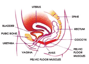
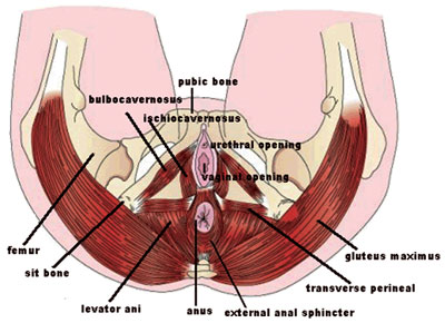
The pelvic floor is a muscular sling that supports the abdomino- pelvic organs, including the bladder in front, the uterus centrally and the lower bowel and rectum behind. Through the pelvic floor muscles,pass the tubular outlet structures of the urethra (the bladder outlet), the vagina (the front passage), and rectum(the back passage). Your pelvic floor muscles help you to control your bladder and bowel. They also help sexual function. It is vital to keep your pelvic floor muscles strong.
Women of all ages need to have strong pelvic floor muscles.
Pelvic floor muscles can be made weaker by:
- Not keeping them active
- Not being pregnant and having babies
- Constipation
- Being overweight
- Heavy lifting
- Chronic coughing (such as smoker’s cough, asthma or bronchitis)
- Growing older
Women with stress incontinence
That is women who wet themselves when they cough, sneeze or are active- will find pelvic floor muscle training can help in getting over this problem.
For pregnant women
Pelvic Floor muscle training will help the body cope with the growing weight of the baby. Healthy, fit muscles before the baby is born will mend more easily after the birth.

After the birth of your baby
You should begin pelvic floor muscle training as soon as you can. Always try to brace your pelvic floor muscles (squeeze up and hold) when you remember during the day and each time you lift your baby.

As women grow older, the pelvic floor muscles need to stay strong because hormone changes after menopause can affect bladder control. As well as this, the pelvic floor muscles change and get weak. A pelvic floor muscle training plan can help to lessen the effects of menopause on pelvic support and bladder control
3D/4D Pelvic Floor Ultrasound
Pelvic Floor ultrasound
3D/4D Pelvic floor ultrasound is a simple study that can obtain images comparable to a MRI ,with the advantage of watching the way the pelvic floor muscles are moving in real time.
The images obtained are from a transperineal approach, meaning the ultrasound probe (transducer) is placed externally on the labia. The data obtained can then be manipulated to view the pelvic floor muscles and organs in three planes and in multiple slices similar to a CT scan.
How it helps to diagnose Prolapse?
Although prolapse is very often diagnosed clinically, pelvic floor ultrasound can help quantifiy the degree of prolapse of either the bladder, uterus or rectum. It is especially useful in differentiating between different forms of posterior compartment problems. For example, ultrasound can help differentiate a rectocele (descent of the rectum) from a enterocele (descent of the small bowel).
How it helps with symptoms of urinary incontinence ?
With ultrasound we can look at bladder neck mobility. Over-rotation often indicates stress incontinence. We can also look for funneling of the bladder neck which is often associated with leakage or urge incontinence.
How ultrasound can help tell you if you are doing your pelvic floor exercises correclty
Ultrasound can give you dynamic feedback on your pelvic floor muscle strength. By watching the monitor with a trained sonographer you will be able to see the pelvic floor muscles contracting and relaxing as you squeeze.


